
When You Have an Extra Tooth in the Front of Your Smile
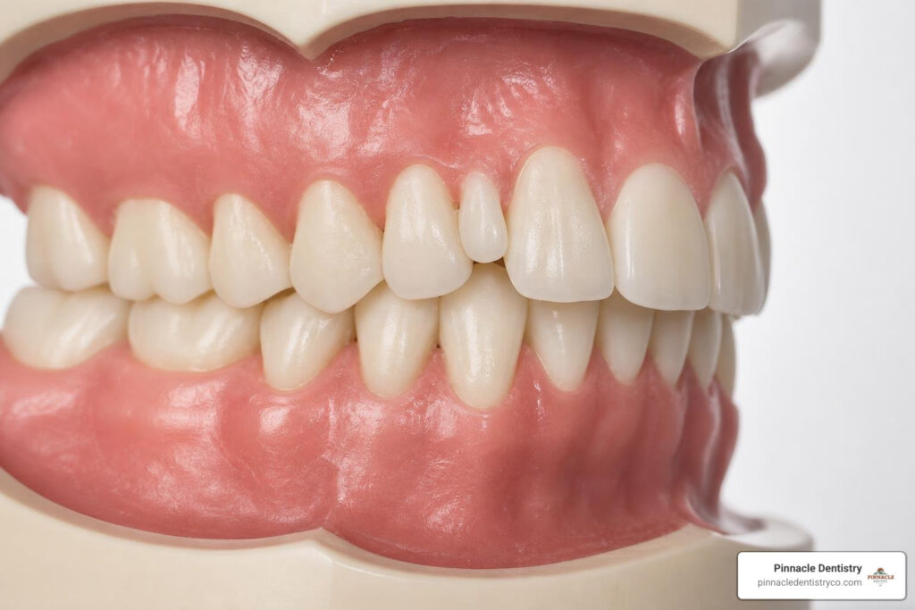
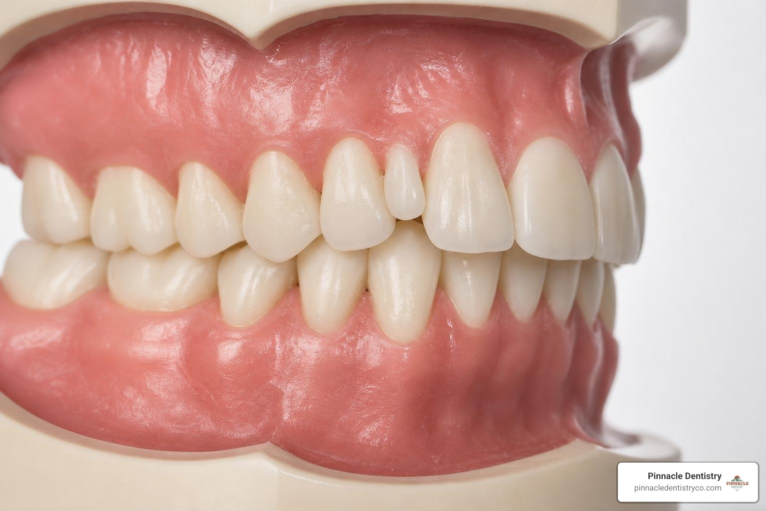
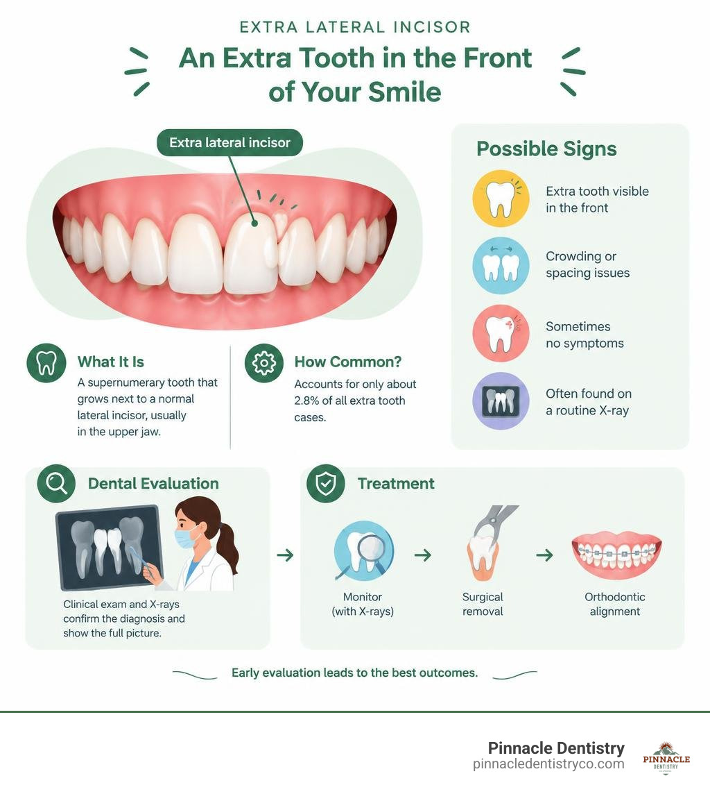
An extra lateral incisor is a supernumerary, or extra, tooth that grows next to a normal lateral incisor, the small tooth between a front tooth and canine. Here is a quick overview of what you need to know:
- What it is: An extra tooth that develops alongside a normal lateral incisor, most often in the upper jaw
- How common: Supernumerary lateral incisors account for only about 2.8% of all extra tooth cases, making them quite rare
- What causes it: Overactivity of the dental lamina, the tissue responsible for forming teeth, during development, often with a genetic link
- Main risks: Crowding, blocked eruption of permanent teeth, root resorption, and in some cases cyst formation
- Treatment: Ranges from monitoring with regular X-rays to surgical removal followed by orthodontic alignment
Most people with an extra lateral incisor do not realize it is there until a routine dental X-ray reveals it. In fact, about 75% of supernumerary lateral incisors cause no symptoms at all. That said, leaving one untreated without professional guidance can lead to serious complications over time, including damage to the roots of neighboring teeth.
As a parent, discovering that your child has an unexpected extra tooth can feel alarming. The good news is that with the right diagnosis and a clear treatment plan, this condition is very manageable.
At Pinnacle Dentistry, our experienced dental team provides personalized evaluations for complex concerns involving an extra lateral incisor in Colorado Springs, CO. In the sections below, we explain how this condition is classified and what your treatment options may look like.
What is an Extra Lateral Incisor and How is it Classified?
To understand how an extra lateral incisor affects your mouth, we first have to look at how dentists classify extra teeth. In the dental world, having more than the standard number of teeth, 20 primary baby teeth or 32 permanent adult teeth, is known as hyperdontia. These extra teeth are clinically termed supernumerary teeth.
When a supernumerary tooth develops in the front of the mouth, it directly affects the anterior teeth, which are the six teeth at the front of each jaw responsible for smile aesthetics and initial biting function. A scientific review of supernumerary lateral incisors explains that these anomalies are classified based on their shape, size, and location in the dental arch.
Understanding Supplemental vs. Rudimentary Extra Lateral Incisor Types
When we diagnose a supernumerary tooth, we look closely at its physical shape, or morphology. This helps us place it into one of two primary categories:
- Supplemental (Eumorphic) Teeth: These are duplicate teeth that closely mimic the normal shape, size, and anatomy of a standard tooth. A supplemental lateral incisor can look almost identical to a natural lateral incisor, making professional evaluation important.
- Rudimentary (Dysmorphic) Teeth: These teeth look abnormal and are typically smaller than standard teeth. They can be conical, or peg-shaped, tuberculate, with multiple small cusps, or molariform, resembling tiny molars.
While most supernumerary teeth in the mouth are rudimentary and peg-shaped, the maxillary lateral incisor is unusual because an extra one may be supplemental and closely resemble its neighboring tooth. The dental team must carefully determine which tooth offers the strongest long-term prognosis before recommending treatment.
Location and Position Classifications
Supernumerary teeth are also classified by where they appear in the jaw:
- Mesiodens: The most common type of extra tooth, which grows directly between the two central front teeth at the midline.
- Paramolars: Extra teeth that grow on the cheek or tongue side of standard molars.
- Distomolars: Extra teeth that grow behind the very last molars in the back of the mouth.
- Premaxillary Supernumeraries: Teeth that develop in the premaxilla, the specialized front section of the upper jaw. This is where extra lateral incisors reside.
Because the premaxilla has a different embryonic development pathway than the rest of the upper jaw, it is a common site for supernumerary anomalies.
Prevalence and Causes of Supernumerary Anterior Teeth
Hyperdontia is a relatively uncommon condition, affecting between 1% and 3% of individuals, with some global studies showing a range up to 5.3% in permanent adult teeth. It is much rarer in baby teeth, occurring in only about 0.3% to 0.8% of children. Interestingly, hyperdontia in adult teeth is more common in males than females.
While extra teeth can grow anywhere, they are far more common in the upper jaw, or maxilla, than the lower jaw, or mandible. Among these, supernumerary lateral incisors and canines are exceptionally rare, representing only about 2.8% of recorded hyperdontia cases.
The Rarity of Bilateral Extra Lateral Incisor Cases
If finding a single extra lateral incisor is rare, finding them on both sides of the mouth, known as bilateral cases, is even less common. Symmetrical, bilateral supplemental lateral incisors represent a small share of supernumerary cases.
Published case reports show that bilateral supplemental lateral incisors can remain unnoticed until crowding or orthodontic concerns lead to a detailed radiographic evaluation. These cases reinforce the importance of evaluating the full position and root development of each tooth before treatment.
Genetic and Environmental Triggers
Why do some people grow extra teeth? While the exact cause remains under scientific discussion, three primary theories explain the development of supernumerary teeth:
- The Dental Lamina Hyperactivity Theory: This is the most widely accepted explanation. During early fetal development, a band of tissue called the dental lamina forms tooth buds. If localized cells in this tissue become hyperactive, they can produce additional tooth buds, leading to extra teeth.
- The Tooth Bud Dichotomy Theory: This theory suggests that a single normal tooth bud accidentally splits into two equal or unequal parts during early development, creating a twin tooth.
- The Atavism Hypothesis: Some evolutionary biologists suggest that extra teeth are a genetic throwback to ancestors who possessed more teeth in their dental formulas.
Genetics may play a role, and extra lateral incisors can occur among family members without being associated with a systemic disorder. When a patient presents with multiple supernumerary teeth, however, the dental team may evaluate for associated conditions such as cleidocranial dysplasia, Gardner syndrome, or cleft lip and palate.
Symptoms and Complications of Having an Extra Tooth
While many supernumerary lateral incisors do not cause immediate symptoms, they can act as silent disruptors in the mouth. As the extra tooth tries to find space in an already crowded jaw, it can trigger several physical and aesthetic issues.
Impact on Adjacent Permanent Teeth
An extra lateral incisor can severely affect the surrounding permanent teeth. Because there is simply not enough room in the premaxilla for an extra tooth, it may block the natural eruption of adjacent teeth. In some cases, supernumerary teeth contribute to delayed or completely blocked eruption of permanent central incisors or canines.
Additionally, pressure from an extra tooth can cause neighboring teeth to rotate, tilt, or drift out of position. This can lead to a noticeable midline deviation, where the center line of the front teeth does not align with the center of the face, or contribute to bite misalignment.
Long-Term Pathological Risks
Leaving an extra tooth unmanaged can lead to hidden, progressive damage beneath the gums:
- Root Resorption: Pressure from an erupting or impacted extra tooth against the roots of adjacent healthy teeth can cause the body to break down those roots. This is known as root resorption and, if left unchecked, can weaken neighboring teeth.
- Dentigerous Cysts: If the extra tooth remains impacted under the gums, the fluid-filled sac surrounding the developing tooth can expand and form a dentigerous cyst. These cysts can damage surrounding jawbone and nearby teeth.
- Oral Hygiene Challenges: Severe crowding caused by an extra tooth can make brushing and flossing difficult. This increases the localized risk of tooth decay, plaque buildup, and periodontal disease.
How Dentists Diagnose and Evaluate Supernumerary Teeth
Because many extra teeth remain asymptomatic and trapped beneath the gumline, they are typically discovered during routine diagnostic examinations at Pinnacle Dentistry. Early detection is key, which is why we recommend children have their first comprehensive dental screening by age seven, when their permanent front teeth begin to erupt.
Clinical Examination and the Flossing Test
Our diagnostic process begins with a detailed physical evaluation. We count the teeth, inspect the symmetry of the dental arch, and look for signs of crowding or delayed eruption.
If we suspect a supplemental tooth, we might perform a clinical “flossing test.” By sliding dental floss between the teeth, we can check for tight, abnormal contact points or detect whether two highly similar teeth are completely separate or fused together.
Advanced Radiographic Imaging
Standard visual exams are not enough to see what is happening beneath the bone. We rely on advanced dental imaging to map out the exact position of the extra tooth:
- Panoramic X-rays: These provide a broad, two-dimensional view of your entire mouth, showing us the general layout of erupted and unerupted teeth.
- Periapical and Occlusal Films: These targeted X-rays offer high-detail views of the roots and surrounding bone structure in the front of the mouth.
- Cone-Beam Computed Tomography (CBCT): This is the gold standard for diagnosing impacted supernumerary teeth. CBCT provides a highly detailed 3D digital model of your jaw, allowing us to see the exact relationship between the extra tooth, neighboring roots, and local nerves. This level of precision is vital for planning safe, minimally invasive treatments.
Treatment Options and Clinical Solutions for Extra Teeth
Once we have diagnosed an extra lateral incisor, we develop a highly personalized treatment plan. There is no one-size-fits-all approach. The best path forward depends on the patient’s age, tooth position, symptoms, and overall orthodontic goals.
When to Choose Extraction vs. Observation
The first major clinical decision we make is whether to remove the extra tooth or simply monitor it.
- Observation: If the extra tooth is completely asymptomatic, fully erupted in acceptable alignment, and causing no damage to adjacent teeth or gums, we may recommend leaving it alone. We will monitor it closely during routine visits with periodic radiographs.
- Strategic Extraction: Extraction is indicated if the tooth is causing crowding, blocking other teeth, causing root resorption, or preventing proper hygiene.
If we must extract a supplemental tooth that looks identical to the natural one, we evaluate which tooth is more displaced from the natural arch, which has the healthier root structure, and which has better periodontal support. We prioritize preserving the tooth with the strongest long-term prognosis.
Timing is also critical. In pediatric cases, removing an obstructing extra tooth early can give the natural permanent teeth a better opportunity to erupt. Our guide to restorative dentistry explains how dental care can support long-term function after treatment is complete.
Restorative and Cosmetic Solutions After Extraction
After the extra tooth is removed, our team may coordinate orthodontic care to close gaps and align the remaining teeth. If the tooth or surrounding structure needs repair after treatment, restorative dentistry care can help rebuild comfortable, functional results.
Once teeth are in their proper positions, dental bonding may be used to refine a slightly smaller or peg-shaped lateral incisor. In appropriate cases, veneers may also help improve the tooth’s shape and appearance.
If a natural tooth is lost because of severe root resorption, the team can discuss replacement options after a full evaluation. For more context on available solutions, see our overview of the types of dental implants in Colorado Springs.
Frequently Asked Questions about Extra Lateral Incisors
What is an extra lateral incisor?
An extra lateral incisor is a supernumerary tooth that develops in the anterior maxilla, the upper front jaw. It grows alongside your standard lateral incisors, which are the teeth located between your central front teeth and your canines. It often presents as a duplicate tooth that looks almost identical to your natural incisor.
How rare is a supernumerary lateral incisor?
Supernumerary lateral incisors are highly uncommon, making up only about 2.8% of all extra tooth cases. Symmetrical, bilateral cases, where an extra tooth appears on both sides of the front arch, are even scarcer, representing less than 10% of these rare anomalies.
What causes an extra lateral incisor to grow?
While the exact cause is not fully understood, the most widely accepted theory is the local hyperactivity of the dental lamina, the tissue responsible for tooth development. Genetic factors, tooth bud splitting, and evolutionary reversion are also believed to play significant roles.
What are the symptoms of an extra lateral incisor?
Roughly 75% of symmetrical supernumerary lateral incisors do not cause immediate symptoms. However, when symptoms do occur, they typically present as dental crowding, localized pain, tooth displacement, or delayed eruption of adjacent permanent teeth.
How is a supernumerary lateral incisor diagnosed?
Because these teeth are often hidden beneath the gums, they are primarily diagnosed through routine clinical examinations and advanced dental imaging. Dentists use panoramic X-rays, detailed periapical films, and 3D CBCT scans to locate and evaluate the tooth.
Does an extra lateral incisor always need to be removed?
No, extraction is not always mandatory. If the extra tooth is healthy, asymptomatic, and not causing crowding or damage to neighboring teeth, we may recommend long-term observation. However, extraction is necessary if it causes functional, aesthetic, or pathological issues.
Can an extra lateral incisor affect the eruption of other teeth?
Yes, this is one of the most common complications. An extra tooth in the premaxilla frequently blocks the eruption pathway of permanent central incisors and canines, causing them to become impacted or erupt in highly abnormal positions.
What happens if an extra lateral incisor is left untreated?
If left unmanaged, a problematic extra tooth can cause root resorption of adjacent healthy teeth, lead to the formation of painful dentigerous cysts, increase the risk of decay and gum disease due to hygiene challenges, and cause severe bite misalignment.
How does an extra lateral incisor impact orthodontic treatment?
An extra tooth complicates orthodontic mechanics by consuming valuable space in the dental arch. Managing it requires careful coordination, often involving strategic extraction followed by orthodontic appliances to align the remaining teeth and correct the bite.
Are extra lateral incisors hereditary?
Yes, there is a strong genetic link. Studies have documented the occurrence of non-syndromic extra lateral incisors in siblings, suggesting that the trait can be inherited. However, they can also occur as isolated, random developmental anomalies.
Conclusion
Discovering an extra lateral incisor can feel overwhelming, but modern dentistry offers gentle, effective ways to manage it. Whether your case requires periodic observation, strategic extraction, or orthodontic and cosmetic rehabilitation, the goal is to protect long-term oral health and restore confidence.
At Pinnacle Dentistry, our highly trained team provides personalized, high-quality care to families throughout Colorado Springs, CO, and El Paso County, CO. We combine advanced diagnostic technology with a warm, patient-first approach to support your comfort at every step.
If you suspect that you or your child may have an extra tooth, or if you are due for a comprehensive diagnostic checkup, our team is here to help. A personalized evaluation can clarify the position of the tooth, its effect on neighboring teeth, and the most appropriate next step.
Ready to take the next step toward a healthy, beautifully aligned smile? Contact our extra tooth treatment specialists at Pinnacle Dentistry to schedule your personalized consultation.